CT Scanner
What is CT scanning?
Computerised tomography (CT) is a test that uses X-ray equipment and computer software to create detailed 2d-pictures of the inside of your body. It takes images of 'slices' of your body (2-d images) and then some sophistaicated software can reassemble them into a 3-d image in the latest machines.
A CT scan results in your body being subjected to a dose of radiation that is over 100 times that for a simple X-ray as it takes hundreds of 'shots'.
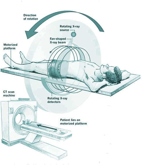
 The CT scanner is a ring-shaped machine. Inside the ring is an X-ray tube that produces a narrow, fan-shaped, monochromatic beam of X-rays. These are fired through your body towards an array of detectors on the opposite side of the ring (you are not expected to learn about the construction or operation of these for A2 AQA A level). The tube and the detectors rotate around your body as you lie flat, creating individual pictures that are cross-sections - slices - through your body.The fan allows several 'slices' to be done at one time - reducing the time of the scan.
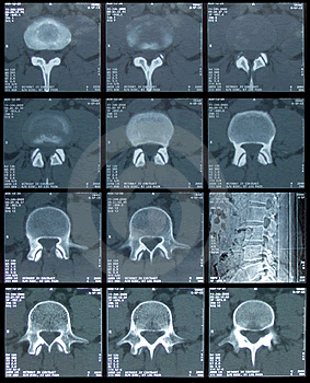 The images from CT are produced from data of X-ray intensity - this is now usually done by computer software (but older systems may produce' film slices'. The images are usually monochrome (black, white and grey), as with ordninary X-rays, but they can be colour coded (in new sophistacated machines) by the doctor to highlight different densities of tissue. Computer software is used to join 'the slices' together to give three-dimensional views. This means that a CT scan allows doctors to see the height, width and depth of something inside the body.
The images are usually stored in a digital format and can then be shown on a computer screen. (Older systems may produce films which would be put up on an illuminated screen - but this is not on the syllabus!).
The amount of time you spend in the scanner and the preparations made beforehand vary depending on which part of your body is being scanned and which conditions your doctors are aiming to diagnose. Most machines now use a technology called 'multislice' scanning (hence the 'fan shaped' narrow beam), and can carry out detailed scans in a matter of seconds.
CT scans are used to diagnose and monitor many health conditions including cancer. They can also be used to provide views of your body so that another procedure or treatment can take place, for example, taking a biopsy (small sample of tissue).
What are the alternatives to a CT scan?
Alternative imaging procedures include ultrasound and magnetic resonance imaging (MRI). There may be other ways of looking inside the body, such as an endoscopy or surgery. Your doctor will explain the benefits and risks of having a CT scan and discuss with you which procedure is most suitable for you.
Having a scan
A radiographer (a health professional trained to perform imaging procedures) will operate the scanning equipment. You must tell your radiographer about any medicines you're taking and if you have any allergies, glaucoma or heart disease.
If you're a woman of childbearing age, you will be asked if you're pregnant. This test isn't recommended for pregnant women, unless there is an urgent medical reason. For example, if there is a risk that you may have a blood clot on your lung, you may need to have a CT scan to diagnose this. You should always tell your radiographer if you could be pregnant.
 Contrast medium
Depending on the part of your body being examined, a contrast medium may be used to make some tissues show up more clearly. This is harmless to your body and you will pass it out in your urine. However, rarely, you may be allergic to the chemical that is used so your radiographer will ask you if you have previously had an allergic reaction to this. He or she may also ask if you get asthma or have an allergy to shellfish because these may make it a little more likely that you will have side-effects from the 'dye'.
The dye is injected into a vein usually in your hand or arm.
If you're having a scan of your stomach or bowels, you may be given a special drink before the scan. This allows your bowels to show up more clearly. For some scans of the colon and rectum, dye and/or air may be inserted into your back passage. Your radiographer should explain what is involved with the examination and ensure that you're happy to proceed.
About the procedure
The time in the scanner takes from 10 to 45 minutes depending on what type of scan you're having. If an additional procedure or treatment is planned, it may take longer.
You will be asked to remove your clothing and put on a gown. For head scans you may be asked to remove contact lenses, dentures, hair clips and hearing aids. For other scans you may be asked to take off your jewellery and wristwatch.
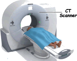
The scanner is a large, ring-shaped machine with a hole in the centre. Only the part of your body inside the ring can be scanned. You will be asked to lie on a table that can slide in or out of the ring.
Your radiographer will position the table so that the part of your body to be scanned is in the centre of the scanner. The table will move backwards or forwards slowly. The X-ray unit inside the ring will rotate around you as the table moves - this creates a new scan each time it goes round. You may be asked to inhale, exhale or hold your breath at certain points during the scan. For the rest of the time, it's important to lie very still.
Your radiographer will operate the scanner from a control room behind a thick lead glass window. He or she will be able to see, hear and speak to you during the procedure.
If you get claustrophobic, it's a good idea to mention this to your doctor or radiographer beforehand.
What to expect afterwards
When the scan is complete, the table is moved back out of the scanner and you will be helped down. When you feel ready, you can go home.
If you had a contrast injection, wait an hour before driving. Try to keep well hydrated for the next 24 hours. An extra glass of water every hour may be helpful.
If you're breastfeeding, it's recommended that you wait 24 hours after having a contrast injection before you breastfeed. You may need to express and discard your milk for 24 hours. Please ask your doctor or radiographer for advice.
Results
A radiologist, a doctor who specialises in using imaging methods to diagnose medical conditions, will look at the images. A report will be sent to the doctor who requested your test - this can take several days.
What are the risks?
CT scans are commonly performed and generally safe. In order to make an informed decision and give your consent, you need to be aware of the possible side-effects and the risk of complications for this procedure.
You will be exposed to X-ray radiation. This can increase your risk of getting cancer - more exposure - greater increase of the probability that the ionising radiation can cause mutations in the DNA of cells leading to cancer. However, any risk of having a CT scan is generally heavily outweighed by the advantages of having the scan. Your doctor will have weighed up the odds and spoken to you about this.
If you are pregnant
Pregnant women are advised not to have CT scans as there is a risk the ioising radiation may cause some damage to the DNA of the unborn child. If you could be pregnant, please tell your doctor or radiographer.
Side-effects
These are the unwanted but mostly temporary effects of a successful procedure. The contrast injection, if you have one, may give you a warm or flushed feeling, or make you feel that you want to pass urine. These side-effects should last only a minute or two.
Complications
Complications are when problems occur during or after the procedure. Most people aren't affected.
In rare cases, it's possible to have an allergic reaction to the contrast injection. If you have any itching or difficulty breathing, tell your radiographer immediately. Medicines are available to treat any allergic reaction.