 |
3-D Ultrasound
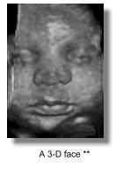
3-dimensional ultrasound has moved out of the research and development stages over the past ten years and is now very much in the News. Faster and more advanced commercial models are coming into the market. The scans requires special probes and software to accumulate and render the images, and the rendering. Scan time has been reduced from minutes to seconds. A good 3D image is often quite impressive and further 2D scans may be extracted from 3D blocks of scanned information. Volumetric measurements are more accurate and both doctors and parents can better appreciate a certain abnormality or the absence of a certain abnormality in a 3D scan than a 2D one and there is the possibility of increasing psychological bonding between the parents and the baby. A large volume of literature and documentation is expected
to come out in the coming years and the diagnosis of congenital anomalies
could receive revived attention. Present evidence has already suggested
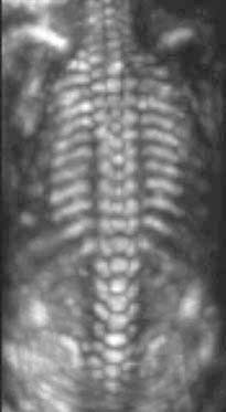
that even small defects such as spina bifida, cleft lips/palate, and
polydactyl may be more lucidly demonstrated. Other more subtle features
such as low-set ears, facial dysmorphia or clubbing of feet can be better
assessed, leading to The study of foetal cardiac malformations is also receiving attention. The ability to obtain a good 3D picture is nevertheless still very much dependent on operator skill, the amount of liquor around the foetus, it's position and the degree of maternal obesity, so that a good image is not always readily obtainable. Experts in this field have not considered that 3D ultrasound will be a mandatory evolution of our conventional 2D scans, rather it is an additional piece of tool like doppler ultrasound. Whether 3D ultrasound will provide unique information or merely supplemental information will only wait to be seen. It's greatest potential is still in research and particularly in the study of foetal embryology.
|
Follow me...
|


Cyberphysics - a web-based teaching aid - for students of physics, their teachers and parents....