
 |
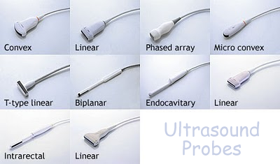
Ultrasound Probes
The thickness, size and location of various soft tissue structures in relation to the origin of the ultrasound beam can be calculated at any point in time using an 'echo technique'. This is called ultrasound scanning. When an ultrasound wave meets an interface of 'differing echogenicity' (big difference in acoustic impedance between the media), the wave is reflected, refracted or absorbed. The reflected sound waves are then processed into an image. Transducer design therefore has to take into account the direction the sound waves need to be transmitted in and the bodily structures that it has to encounter. Ultrasound transducers, often just called probes, therefore come in different shapes and sizes for use in different scanning situations.
For physics medical option examination revision you have to concentrate on the probe itself and the interpretation of scan data. Probes The probe emits ultrasound in rapid pulses, and also acts as a receiver. The ultrasound images can be displayed on an oscilloscope screen or a video monitor (via what is known as a scan converter) and can be recorded on videotape, thermal paper or radiographic film. Pulses of ultrasound can be aimed in a specific direction and obey the laws of geometric optics with regard to reflection, transmission and refraction. The frequency of the ultrasound affects how far it can penetrate into material and therefore the quality of the image produced. A skilled operator will choose the correct frequency for the task in hand. The higher the frequency, the smaller the wavelength of the ultrasonic waves and the less the diffraction. This means that at higher frequency you get greater the detail in the image. The frequency used for a particular application depends on the depth and density of the organ to be imaged. Low density organs near the body's surface (e.g. the eye) can be imaged in more detail than high-density internal structures (e.g. a baby in the womb) because higher frequency ultrasonic waves can be used for lower density organs near the surface. Such high frequency ultrasonic waves would be mostly absorbed by tissue they have to pass through before they reach the internal organ. High frequency ultrasound waves
Low frequency ultrasound waves
See here for details of the factors that affect how ultrasound is reflected. There are two main types of scan that are performed. In order to perform an A-scan a transducer with a single crystal can be used - called a single transducer probe - but for a B-scan a multi=transducer probe is needed. Click on the links below for more detail on the scan methods:
|
Follow me...
|
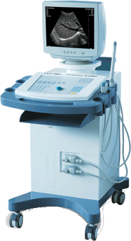 An ultrasound scanning system comprises of probes, a scan converter and a monitor. The scan converter enables the operator to set the frequency and beam spread. Focusing and aperture control technology are often employed to narrow the beam along it's entire path to achieve maximum right-and-left (lateral) resolution. The converter also often allows for the output to be


Cyberphysics - a web-based teaching aid - for students of physics, their teachers and parents....