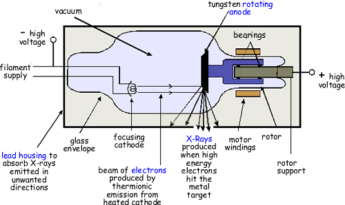
The X-Ray Tube
The voltage to the tube is supplied by a circuit composing of mains electricity and a step up transformer. A high voltage is needed to produce the kinetic energy required of the electrons to and a relatively lower one is used for the filament cathode. This is achieved by a potential divider circuit.
Electrons are produced
by thermionic emission in the cathode. This is heated by a relatively
low voltage supply.
At a cathode current of 100 mA, for example, 6 x
1017 electrons will travel from the cathode to the anode of the
X-ray tube every second.They are accelerated
from the cathode to anode across an alternating high voltage - they will therefore only be attracted in half of the cycle. As the kinetic energy
of the electrons increases, both the intensity (number of x-rays) and
the energy (their ability to penetrate) of the X-rays produced are increased.
When these electrons
bombard on the heavy metal atoms of the target, they interact with these
atoms and transfer their kinetic energy to the target. These interactions
occur within a very small depth of penetration into the target. As they
occur, the electrons slow down (brake!) and finally come nearly to rest,
at which time they can be conducted through the x-ray anode assembly
and out into the associated electronic circuitry.
The interactions
result in the conversion of kinetic energy into thermal energy and electromagnetic
energy in the form of X-rays.
Most of
the the kinetic energy is converted into heat. The electrons interact
with the outer-shell electrons of the target atoms but do not transfer
sufficient energy to these outer-shell electrons to ionize them. Rather,
the outer-shell electrons are simply raised to an excited, or higher,
energy level. The outer-shell electrons immediately drop back to their
normal energy state with the emission of infrared radiation. The constant
excitation and restabilization of outer-shell electrons is responsible
for the heat generated in the anodes of X-ray tubes.
Generally, more than
99% of the kinetic energy of projectile electrons is converted to thermal
energy, leaving less than 1% available for the production of X-radiation.
In this sense,the X-ray machine is a very inefficient apparatus.
The production of
heat in the anode increases directly with increasing tube current. Doubling
the tube current doubles the quantity of heat produced.
Heat production
also varies almost directly with varying the high tension voltage too.
The efficiency of
X-ray production is independent of the tube current. Regardless of what
mA is selected, the efficiency of X-ray production remains constant.
The efficiency of X-ray production increases with increasing projectile-electron
energy. At 60 keV, only 0.5% of the electron kinetic energy is converted
to X-rays; at 120 MeV, it is 70%.
Target
material
The anode is made to rotate at steady speed so the point of impact continually changes to prevent overheating. But it stillneeds to have:
- a high Z (proton number) so that transitions of high
enough energy to emit X-ray radiation are possible
- a high melting point because so much heat energy is
produced.
Tungsten is ideal (Molybdenum for softer X-rays needed for breast X-rays)
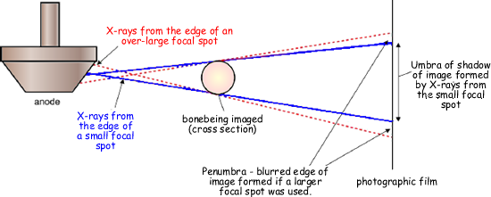
Focal Spot
The area of the anode from which X-rays are emitted is referred to as the focal spot. This must be as small as possible otherwise features in the image would be blurred instead of being sharp. The anode surface is at an angle of about 70° to the electron beam so that the X-rays effectively originate from a much smaller area than the impact area of the beam.
The Options....
Action |
Effect |
Graph
of Intensity against X-Ray photon energy |
Clarity
of image |
| Increasing
the tube voltage |
Increasing
the high p.d. that is used to accelerate the electrons will give
the average electron more energy when it hits the target |
Shape of
spectrum spreads out to encompass higher energies
range is
increased
Characteristics
in the same place (natch!!)
area under
the curve increases |
Too high
an energy of X-ray will penetrate too well to give good definition
- if they all get through - no shadow - picture!
60-125 kV is usually employed - giving energy of about 30 keV |
AC/DC voltage
(AC necessary
to get higher voltages - can use transformers! DC acquired by electronic
rectification and 'smoothing' circuitry) |
Electrons
produced by thermionic emission only accelerated across half of
the time! |
graphs
for both are the same except the DC one is double the intensity
throughout (only accelerated across to target on half of the
wave). |
|
| Increasing
the tube current (low voltage one!) |
Increases
the rate of thermionic emission - more electrons hit the target -
more X-rays produced. |
Shape of
spectrum remains the same
range is
the same
Characteristics
in the same place (natch!!)
area under
the curve increases |
Overall
increase of exposure of film
but bigger
dose to patient!
more heating
of the target |
| Increasing
exposure time |
|
|
Overall
increase of exposure of film
but bigger
dose to patient!
more heating
of the target
risk of
blur due to movement of patient - big problem with organs that
cannot be constrained. |
| Changing
Target Material |
An increase
in Z (proton number) will increase the probability of electron interactions
of enough energy to produce X-rays - so more X-rays will be produced. |
The Characteristic
peak positions will change - Ks will shift towards higher energies
(these depend on the target material!).
range is
the same
area under
the curve increases |
allows choice
of X-ray energies that give best difference in attenuation for
the part to viewed.
soft X-rays
are needed for soft tissue - harder ones for bone. |
| Using
a filter (material placed in the X-ray beam path) |
Absorbs
mainly lower energy X-rays - and produces a 'harder' more penetrating
beam) |
area under
the curve is smaller (as some of the X-rays have been absorbed).
Shape changes
as mainly X-rays are reduced from the lower energy values.
range is
smaller - but high energy the same.
Characteristics
in the same place (natch!!) |
reduces
unwanted X-rays and therefore the scatter due to them - better
contrast
|
| Reducing
beam size |
|
|
less scatter
- better contrast - especially if a collimator is used (lead
grid that only allows X-rays in a particular direction to get
through. |
| Focal
spot size |
|
|
Small focal
spot produces sharp images
BUT also
intense heating of target |
| Artificial
Contrast Media |
See Barium Meal and Enema |
|
Clearly outlines the inner surface of internal bodily organs
by coating them in a radio-opaque material - barium sulphate.
|
| Intensifying
Screens |
Decreases
the required exposure time. |
|
- Make image clearer with a lower X-ray dose
|
| Detectors |
photographic film |
|
|