
 |
Magnetic Resonance Imaging USES Magnetic resonance imaging (MRI) can be used to look at almost any part of the body. It is most often used to study:
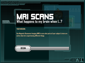
Functional MRI Functional MRI is a type of MRI that allows you to see which parts of your brain are active when you perform different tasks or feel certain emotions and sensations. Rather than taking a single scan, functional MRI takes repeated scans, usually one a second. These are used to track the movement of blood through the brain.
By studying the movement of blood, it is possible to tell which sections of the brain are particularly active in real time and to see how brain activity responds to outside events and activities. For example, a volunteer may be asked to solve a problem or to remember a short phrase. Functional MRI will then be used to determine which parts of the brain are active during these tasks. fMRI mapping of the brain is used to find out how the brain carries out mental tasks and what parts of the brain are responsible for different brain disorders. Functional MRI is a relatively new technique, but it has been used by a number of specialists to help plan complex brain surgery. Click on the image on the right. It will open a page that shows you the types of image produced when you experience different emotions.
|
Follow me...
|


Cyberphysics - a web-based teaching aid - for students of physics, their teachers and parents....