The action potential of heart muscle
 Each time the heart beats, its electrical potential changes by more than 100 mV as the muscles of the heart contract and relax in a sequence that forces blood from the two atrial chambers of the heart into the corresponding ventricles and then out to the arteries.
This sequence is due to nerve cells that conduct electrical signals generated at the sino-atrial (SA) node in the right atrium. These nerve cells extend from the SA node across the surface of the heart, making the muscles of the atrial chambers contract and stimulating the atrio-ventricular (AV) node. This relays the electrical signals to further nerve cells which make the ventricles contract.
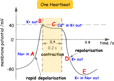
The diagram shows how the potential of the heart changes each time the heart beats.
An electrical impulse from the SA node depolarises then repolarises the nerves of the atria, making the atria muscles contract then relax. The impulse reaches the AV node which, after a delay, then stimulates the nerves of the ventricles, making the muscles of the ventricles relax.
Stage A - rapid depolarisation phase
Once the cell is electrically stimulated (typically by an electric current from an adjacent cell), it begins a sequence of actions involving the influx and efflux of cations and anions that together produce the action potential of the cell. The slope of the graph of this phase represents the maximum rate of potential change and is known as dV/dtmax. In heart muscle cells, this gradient is directly proportional to the net ionic current.
This phase is due to the opening of the fast Na+ channels causing a rapid increase in the membrane conductance to Na+ and thus a rapid influx of Na+ ions into the cell; a Na+ current flows. The ability of the cell to open the fast Na+ channels during this phase is related to the membrane potential at the moment of excitation. If the membrane potential is at its baseline (about -85 mV), all the fast Na+ channels are closed, and excitation will open them all, causing a large influx of Na+ ions. If, however, the membrane potential is less negative, some of the fast Na+ channels will be in an inactivated state insensitive to opening, thus causing a lesser response to excitation of the cell membrane and a lower Vmax. For this reason, if the resting membrane potential becomes too positive, the cell may not be excitable, and conduction through the heart may be delayed, increasing the risk for arrhythmias.
Stage B (start of contraction)
This phase of the myocyte action potential occurs with the inactivation of the fast Na+ channels. The transient net outward current causing the small downward deflection of the action potential is due to the movement of K+ .
Stage C (contraction)
This "plateau" phase of the cardiac action potential, is sustained by a balance between inward movement of Ca2+ through L-type calcium channels and outward movement of K+ through the slow delayed rectifier potassium channels. The sodium-calcium exchanger current and the sodium/potassium pump current, also play minor roles during this phase. It sustains muscle contraction. During the plateau phase there is reduced potassium ion permeability.
Stage D - the "rapid repolarization" phase
During this phase of the action potential, the Ca2+ channels close, while the slow delayed rectifier K+ channels are still open. This ensures a net outward current, corresponding to negative change in membrane potential, thus allowing more types of K+ channels to open. These are primarily the rapid delayed rectifier K+ channels and the inwardly rectifying K+ current. This net outward, positive current (equal to loss of positive charge from the cell) causes the cell to repolarize. The delayed rectifier K+ channels close when the membrane potential is restored to about -80 to -85 mV.
Stage E - the resting membrane phase
The membrane potential when the cell is not being stimulated. In the ventricular myocardium it is about -85 to -95 mV (look at the lowest value on the diagram above - it dips below -80 mV).
This potential is determined by the selective permeability of the cell membrane to various ions. The membrane is most permeable to K+ and relatively impermeable to other ions. The resting membrane potential is therefore dominated by the K+ equilibrium potential according to the K+ gradient across the cell membrane.