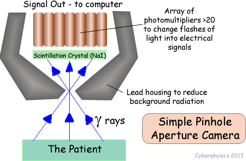
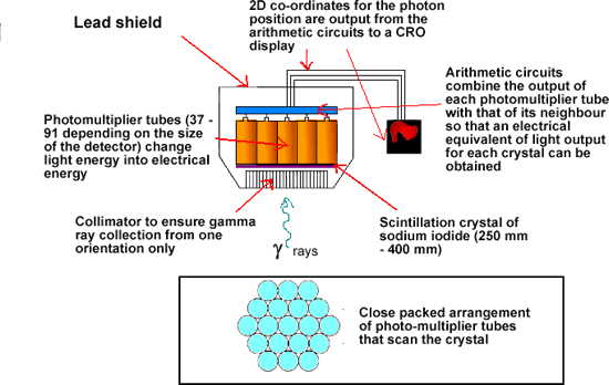
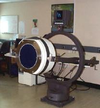
The Gamma Camera

The gamma ray is electromagnetic radiation of very high penetration power. Therefore more rays exit the body and are available for detection than interact with the patient's tissue. These can be detected by a gamma camera and the concentration of radioactive tracer in various parts of the body can be ascertained.

Gamma rays cannot be focused by refraction therefore a lead collimator is used to direct rays from a point on the patient towards a single point on a sodium iodide crystal. The collimator absorbs g-rays emanating from other parts of the body before they activate the crystal.

Pin-hole Aperture Gamma Camera

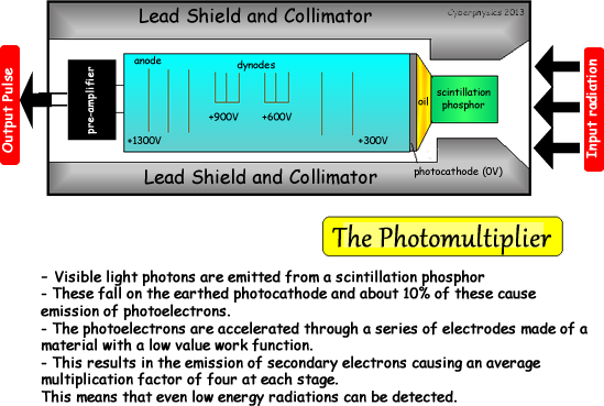
A crystal of sodium iodide fluoresces when a gamma ray interacts with one of its orbital electrons, promoting it to a higher energy level. On its return to ground state a photon is emitted. If this energy is in the visible region a flash of light is seen.

This light is detected using a photomultiplier (see diagram) which is a device in which incident photons create measurable electrical pulses. The device is based on the photoelectric effect (see diagram). It uses large electric fields to accelerate electrons and, through a cascade sequence, amplify the signal.
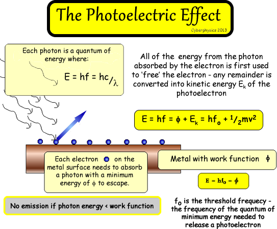
 

Gamma Camera employing parallel hole collimation

  • uses more radiation than pin-hole type without losing resolution
  • is ten times more sensitive when scanning than a pin-hole type


The gamma camera is made to scan the area of interest approximately four hours after the patient has been injected with the radioactive tracer. This gives it time to circulate and accumulate in 'hot-spots' of rapid cell division. The second scan taken within the 24-hour period can then be compared to the first to diagnose suspicion of overactive lymph node activity.

The camera display can be made on a monitor and videoed. Hard copies of the scans can then be taken and compared.

Gamma camera scan of kidneys

The electrical output can be translated into colour coded graphical display using electronic circuitry.

Look at the Heart Gamma Camera Scan graphic - note the use of colour to give instant visual recognition of variations in concentration Scanning is programmed electronically so that the area of interest is looked at. The chemical is chosen because it takes part in a normal biological function of the lymphatic system any slight abnormalities in function can be identified by comparison of the two scans. Current experimentation with a tiny probe containing a miniaturised gamma camera, hopes to combine keyhole surgery with nuclear medicine and hence reduce the risk due to exposure dose of the patient, making more frequent scans a possibility.