ECG - Electrocardiograph
An ECG trace provides important information about the condition of the heart.
The change of electrical potential of the heart is conducted through body fluids and tissue to the skin, causing changes of potential of the order of 1 mV which can be detected by electrodes connected to an amplifier.
An electrocardiograph (ECG) is designed to measure and record the potential difference between two points on the surface of the body. The pd between any two points on the body's surface is depends on:
- the conductivity of the body fluids
- the position of the points on the body
- the action potential of the heart.
A recording of the variation of such a pd with time is called an electrocardiogram or ECG trace.
Detection of biopotentials
Electrodes placed on the surface of the body measure the coordinated activity of a large group of cells and thus register the local action potential, which may be neural or muscular, depending on the electrode locations.
Since body materials, particularly skin, are poor electrical conductors, the selection and preparation of electrode site are most important.
A suitable site must be:
- well-cleaned,
- hair-free and
- rubbed to remove some outer cells.
A conductive, yet non-irritant, electrode paste is rubbed into the site to improve the electrical contact and the applied electrode is then held firmly in place using tape.
Preparing a patient for an ECG
The patient must be striped to the waist to expose the chest (the doctor/nurse should keep the patient's lower half covered as much as possible, they may have seen it all before but should always treat the patient with the utmost respect). The patient's ankles also need to be exposed at this point.
 If using a machine equipped with metallic stickers, it is important that the patient's skin is rubbed with an alcoholic swab before applying them, to ensure good electrical contact is made.
 If the machine is supplied with the older suction cups, then electrolyte spray must be applied to the areas of skin an which electrodes will be placed. Men with very hairy chests may require a gel based electrolyte for adhesion, or in extreme cases, shaving may be needed.
Applying the contacts -
 Accurate chest lead placement is essential for ensuring quality ECG output. Misplacing leads may result in a change in ECG waveform, in turn this may cause the ECG trace to be misinterpreted. This is unacceptable, so chest electrodes must be placed methodically.
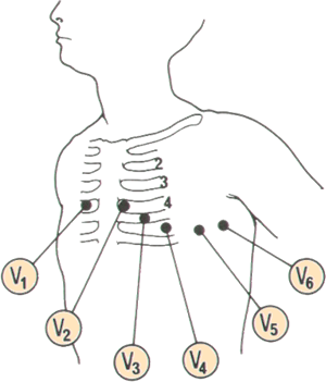
The standard chest lead positions are as follows;
V1 Fourth intercostal space, right sternal edge
V2 Fourth intercostal space, left sternal edge
V4 (Don't worry, not a mistake, place the fourth electrode before the third) Fifth intercostal space in the mid-clavicular line
V3 I could describe this anatomically, but just between you and me, bang it half way between the second and fourth electrodes
V5 Lies on the fifth rib in the anterior axillary line
V6 On an imaginary horizontal line with V5 in the mid axillary line
The rest of the leads:
The four limb leads are easy. One on each arm (go for the wrist, have the metal plate of the clip facing the palm aspect, avoid bony prominences) and one on either ankle. Make sure the right clip is on the right wrist.
The final electrode is the "neutral", it reduces AC interference and in all honesty could be applied to the patient's nose without any adverse effects to the ECG.
See the page on a typical ECG trace.