6.5.1
Using X-rays |
(a) basic structure of an X-ray tube; components – heater (cathode), anode, target metal and high voltage supply |

|
(b) production of X-ray photons from an X-ray tube |
(c) X-ray attenuation mechanisms; simple scatter, photoelectric effect, Compton effect and pair production |
(d) attenuation of X-rays;
I =I0e-μn
where μ is the attenuation (absorption) coefficient |
(e) X-ray imaging with contrast media; barium and iodine |
|
|
(f) computerised axial tomography (CAT) scanning; components – rotating X-tube producing a thin fan-shaped X-ray beam, ring of detectors, computer software and display |
|
|
(g) advantages of a CAT scan over an X-ray image. |
|
|
6.5.2
Diagnostic methods in medicine |
(a) medical tracers; technetium–99m and fluorine–18 |
|
|
(b) gamma camera; components – collimator, scintillator, photomultiplier tubes, computer and display; formation of image |
|
|
(c) diagnosis using gamma camera |
|
|
(d) positron emission tomography (PET) scanner; annihilation of positron–electron pairs; formation of image |
|
Issues raised when equipping a hospital with an expensive scanner. |
(e) diagnosis using PET scanning. |
|
6.5.3
Using ultrasound |
(a) ultrasound; longitudinal wave with frequency greater than 20 kHz
|
|
|
(b) piezoelectric effect; ultrasound transducer as a device that emits and receives ultrasound |
|
(c) ultrasound A-scan and B-scan |
|
(d) acoustic impedance of a medium;
Z = ρc |
|
(e) reflection of ultrasound at a boundary;
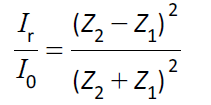
|
|
(f) impedance (acoustic) matching; special gel used in ultrasound scanning |
|
| (g) Doppler effect in ultrasound;
speed of blood in the patient;
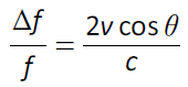
for determining the speed v of blood. |
|