Syllabus Details
Click here to go to the board's book of info on this for teachers |
References |
You should be able to... |
Make use of the past paper questions I have done for you - each question has a solution and comments to help you understand it.
Physics of the eye (past paper questions and solutions) and physics of the ear (past paper questions and solutions) |
| 3.10.1 Physics of the eye |
| 3.10.1.1 Physics of vision |
The eye as an optical refracting system; including ray diagrams of image formation |
Pope pp 28-32
|
- draw ray diagrams to represent the rays being refracted by the optical system (as in Pope) |
|
Sensitivity of the eye
Spectral response as a photodetector |
Pope pp 32-36 |
- diagrams on pages 32, 33 and 35 should be known and understood - they have come up in questions in the past
Maximum refaction occus at the air cornea interface - the lens only provides minor adjustments in focus. - know diagram too!
- depth of focus with a small pupil is large - little adjustment is needed for focusing at differenet object distances - lttle strain when viewing in bright light. The converse is true for a large pupil.- know diagram too!
Rods and cones should be known in detail - learn the graphs on page 35 - you should be able to sketch them from memory with numbers on the axes. |
|
Spatial resolution of the eye
Explanation in terms of the behaviour of rods and cones |
Pope pp 36-32 |
the diagram on page 36 should be known and understood
|
| 3.10.1.2 Defects of vision and their correction using lenses |
Lenses
Properties of converging and diverging lenses; principal focus, focal length and power
Use of the equations:
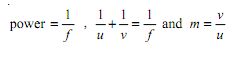
where
f = the focal length (+ve for a convex lens and -ve for a concave one)
u = the distance of the object from the centre (pole) of the lens, called the object distance
v = the distance of the image from the centre (pole) of the lens, called the image distance (if the image is a virtual one this distance will be negative and the image will be on the same side of the lens as the object.
m = magnification
|
|
You should recall that:
 a converging lens makes the rays come to a real focus (cross at a point) it is called a CONVEX lens a converging lens makes the rays come to a real focus (cross at a point) it is called a CONVEX lens
a diverging lens makes the rays appear to be spreading out from a point on the opposite side of the lens (cross at a virtual point) it is called a CONCAVE lens
The principal focus of a convex lens (sometimes called the focal point) it is the point on the principal axis, through which rays of light, travelling near to and parallel to the principal axis, pass after refraction by the lens.
The principal focus of a concave lens (sometimes called the focal point) it is the point on the principal axis, from which rays of light, travelling near to and parallel to the principal axis, apear to diverge from after refraction by the lens. (they can be traced back through this point.
The focal length of a lens is the distance between the pole of the lens and the principal focus. If the lens has a real focus then the focal length is positive. If it has a virtual focus it is negative.
The power of a lens is the reciprocal of the focal length in metres. It is measured in dioptres (D). The sign of the power will be the same as the sign of the focal length. You should be able to do calculations using the equations - care with signs! and units! |
|
Defects of vision: myopia, hypermetropia and astigmatism
Correction of defects of vision using lenses
Ray diagrams and calculations of powers (in dioptres) of correcting lenses for myopia and hypermetropia
The format of prescriptions for astigmatism |
|
Ray diagrams and image formation must be fully mastered.
You should be able to construct the following diagrams
- Convex lens: object at 2F, object between F and 2F, object at F , object between P and F and object beyond 2F
- Concave lens: object at 2F, object between F and 2F, object at F and object between P and F
You MUST draw these properly - construct them! - sketches will not do!
Use a ruler and a sharp pencil.
Three basic rules to construcy an image:
Rays that pass through the pole of the lens are undeviated.
Rays that are travelling parallel to and near to the principal axis pass through the principal focus after refraction by the lens (for a convex lens) and appear to emanate from the principal focus (draw in dotted lines) for a concave lens.
The converse is true, in that rays passing through (or emanating from) the principal focus emerge parallel on the other side of the lens.
|
| 3.10.2 Physics of the ear |
| 3.10.2.1 The ear as a sound detection system |
Simple structure of the ear, transmission processes |
Pope 47-63
Pope 49-51 |
|
| 3.10.2.2 Sensitivity and frequency response |
Production and interpretation of equal loudness curves
Human perception of relative intensity levels and the need for a logarithmic scale to reflect this
Definition of intensity where the threshold of hearing is I0
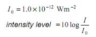
Measurement of sound intensity levels and the use of dB and dBA scales
Relative intensity levels of sounds |
Pope 53-58
Pope 58-61 |
|
| 3.10.2.3 Defects of hearing |
The effect on equal loudness curves and the changes experienced in terms of hearing loss of:
- injury resulting from exposure to excessive noise;
- deterioration with age (excluding physiological changes) |
|
Notes on the Cyberphysics site - see table of problems with hearing |
3.10.3 Biological measurement
Make use of the past paper questions I have done for you - each question has a solution and comments to help you understand it - Heart traces - Q3 and Q4 are no longer on the syllabus - see below
|
| 3.10.3.1 Simple ECG machines and the normal ECG waveform |
Principles of operation for obtaining the ECG waveform;
explanation of the characteristic shape of a normal ECG waveform |
Pope page 70-72 |
|
3.10.4 Non-ionising imaging
Make use of the past paper questions I have done for you - each question has a solution and comments to help you understand it - Endoscope- Ultrasound |
| 3.10.4.1 Ultrasound imaging |
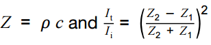
Reflection and transmission characteristics of sound waves at tissue boundaries, acoustic impedance, attenuation
Advantages and disadvantages of ultrasound imaging in comparison with alternatives including safety issues and resolution |
Pope page 117-118
Pope page 130-131 |
Look to the GCSE notes on cyberphysics as well as your text book - also use the extended reading exercise
UCL film on Ultrasound
Pages on colour ultrasound, 3D ultrasound, doppler ultrasound - remember these are 'above and beyond what you need! |
|
Piezoelectric devices: Principles of generation and detection of ultrasound pulses
|
Pope page 118-119 |

The piezo-electric transducer
Probes
Probe use |
|
A-scans and B-scans
Examples of applications
Use of the equations:
|
Pope page 119-125 |
|
| 3.10.4.2 Fibre optics and endoscopy |
Properties of fibre optics and applications in medical physics; including total internal reflection at the core-cladding interface |
Pope pages 109-113 |
Revise AS work on TIR and then see how it is applied here
|
| Endoscopy |
Physical principles of the optical system of a flexible endoscope; the use of coherent and non-coherent fibre bundles; examples of use for internal imaging and related advantages |
Pope pages 109-113 |
Revise AS work on TIR and then see how it is applied here |
| 3.10.4.3 Magnetic resonance (MR) Scanner |
Basic principles of MR scanner:
• cross-section of patient scanned using magnetic fields
• protons initially aligned with spins parallel • spinning hydrogen nuclei (protons) precess about the magnetic field lines of a superconducting magnet
• 'gradient' field coils used to scan cross-section • short radio frequency (RF) pulses cause excitation and change of spin state in successive small regions
• protons excited during the scan emit RF signals as they de-excite
• RF signals detected and the resulting signals are processed by a computer to produce a visual image
Candidates will not be asked about the magnetic fields used in an MR scanner, or about de-excitation
relaxation times. |
Not in Pope |
UCL film on MRI |
3.10.5 X-ray imaging
Make use of the past paper questions I have done for you - each question has a solution and comments to help you understand it - X-rays |
| 3.10.5.1 The physics of diagnostic X-rays |
The physics of diagnostic X-rays |
Pope pages 136 - 160 |
see cyberphysics notes on this topic.
UCL film on X-rays
|
|
Physical principles of the production of X-rays:
- maximum photon energy,
- energy spectrum;
- continuous spectrum and
- characteristic spectrum.
Rotating-anode X-ray tube:
- methods of controlling the beam intensity,
- the photon energy,
- the image sharpness and contrast and
- the patient dose |
Pope pages 151-156 |
Important topic - make sure you know what each part does and why! |
| 3.10.5.2 Image detection and enhancement |
Flat panel (FTP) detector including X-ray scintillator, photodiode pixels, electronic scanning.
Advantages of FTP detector compared with photographic detection.
Contrast enhancement; use of X-ray opaque material as illustrated by the barium meal technique.
Photographic detection with intensifying screen and fluoroscopic image intensification; reasons for using these |
Pope page 149
Pope page
106-107 and 148-151 |
Main reason for development of the intensifying screen etc. is to cut down on the dose of X-rays that has to be delivered. You need to use CALRAD to familiarize yourself with the procedures adopted to do this and the problems that can arise from ionizing radiation exposure.
Cellular damage (within one person) can lead on to systemic (passed on to offspring) damage. See here. |
| 3.10.5.3 Absorption of X-rays |
Exponential attenuation
Linear (attenuation) coefficient m, mass attenuation coefficient mm and half-value thickness
x is thickness and r is density
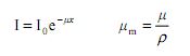
Differential tissue absorption of X-rays excluding details of the absorption processes.
|
Pope pages 142-144
Pope pages 142-147 |
Attenuation is the gradual loss of intensity of any kind of flux as it travels through a medium.
You did something similar with gamma ray absorption - half-value thickness is similar to half life... as you add a half thickness to a material the intensity drops by half again...
Note that you do NOT need details of the attenuation processes! |
| 3.10.5.4 CT scanner |
Basic principles of CT scanner:
- movement of X-ray tube
- narrow, monochromatic
X-ray beam
- array of detectors
- computer used to process the signals and produce
a visual image.
Comparisons will be limited to advantages and disadvantages of image resolution, cost and safety issues. Candidates will not be asked about the construction or operation
of the detectors.
Comparisons of ultrasound, CT and MRI scans; advantages and disadvantages
limited to image resolution, |
Not in Pope |
|
3.10.6 Radionuclide imaging and therapy |
| 3.10.6.1 Imaging techniques |
Use of a gamma-emitting radioisotope as a tracer;
technetium-99m, iodine-131 and indium-111 and their relevant properties.
The properties should include the radiation emitted, the half-life, the energy of the gamma radiation, the ability for it to be labelled with a compound with an affinity for a particular organ. The Molybdenum-Technetium generator, its basic use and importance. PET scans. |
|
UCL film on radiotherapy |
| 3.10.6.2 Half-life |
Physical, biological and effective half-lives;
1/TE = 1/TP + 1/TB
definitions of each term |
|
|
| 3.10.6.3 Gamma camera |
Basic structure and workings of a photomultiplier tube and gamma camera. |
|
|
| 3.10.6.4 Use of high-energy X-rays |
External treatment using high-energy X-rays. Methods used to limit exposure to healthy cells. |
|
|
| 3.10.6.5 Use of radioactive implants |
Internal treatment using beta emitting implants. |
|
|
| 3.10.6.6 Imaging comparisons |
Students will be required to make comparisons between imaging techniques.
Questions will be limited to consideration of image resolution, convenience and safety issues |
|
|
| No longer on the syllabus |
Basic structure of the heart. The heart as a double pump with identified valves
|
Pope page 69/70 |
This section has been removed from the option - but those of you that are going into medical professions might find it interesting or useful when at Uni....
You may wish to make use of the past paper questions I have done for you - each question has a solution and comments to help you understand it - Heart traces |
Electrical signals and their detection
The biological generation and conduction of electrical signals;
methods of detection of electrical signals at the skin surface |
Pope chapter 5 (pp 67-73) |
| Action potential of a nerve cell. The response of the heart to the action potential originating at the sino-atrial node; action potential of the heart muscle. |
Pope page 68-69 |
Simple ECG machines and the normal ECG waveform
Principles of operation for obtaining the ECG waveform;
explanation of the characteristic shape of a normal ECG waveform |
Pope page 70-72 |


 This option offers an opportunity for students with an interest in biological and medical topics to study some of the applications of physical principles and techniques in medicine. It involves detailed learning as well as application of principles to problems and the use and manipulation of equations.
 It builds upon topics covered throughout the course and the studying of this course should be used as an opportunity to practise applying and clarify concepts met in the two years of your studies.

